Is an X-ray when pregnant safe? Yes, generally. Medical X-rays use very low radiation doses, posing minimal risk to your baby, especially with proper shielding. Discuss concerns with your doctor.
By Shubhra Mishra — a mom of two who turned her own confusion during pregnancy into BumpBites, a global mission to make food choices clear, safe, and stress-free for every expecting mother. 💛
Check whether any food is safe during pregnancy with the BumpBites Food Safety Checker.
Download the Complete Pregnancy Food Guide (10,000 Foods) 📘
Instant PDF download • No spam • Trusted by thousands of moms
💡 Your email is 100% safe — no spam ever.
Quick verdict: ⚠️ Talk to your doctor first. A single diagnostic X‑ray is usually safe when shielding is used, but repeated exposure or high‑dose studies should be avoided unless medically essential.
It’s completely normal to wonder about x‑ray when pregnant after a sudden need for imaging or an unexpected appointment. Many expectant parents stare at the waiting‑room poster, replay the moment they learned they’re pregnant, and then worry: “Did that X‑ray hurt my baby?” The good news is that most routine X‑rays deliver a very low dose of radiation, and with proper precautions they are often permissible. In this article we’ll break down the current guidance from ACOG, NHS, and the FDA, explain how radiation dose is measured, and give you a clear, trimester‑by‑trimester safety snapshot. We also aim to empower you with the right questions to ask your healthcare team to ensure the safest possible approach to any necessary imaging.
We’ll also cover how many X‑rays are considered safe, what special steps clinicians take for pregnant patients, and what safer imaging alternatives you can ask for if you’d rather avoid radiation altogether. By the end you’ll know exactly what to discuss with your provider, which imaging methods are low‑risk, and when you can breathe easier about your prenatal care.
Whether you’ve already had a dental X‑ray, are scheduled for a chest exam, or are just curious about the theoretical risks, we’ll walk you through the science in plain language, share practical tips for minimizing exposure, and point you toward safer options when they exist. Remember: you’re not alone—many parents face these questions, and the answers are grounded in solid evidence.
Stage of pregnancy
Verdict
Notes
First trimester (0‑13 weeks)
⚠️ Caution
Use only if medically necessary; fetal organ development is most sensitive.
Second trimester (14‑27 weeks)
✅ Generally safe
Standard diagnostic X‑rays with lead shielding are acceptable.
Third trimester (28‑40 weeks)
✅ Generally safe
Same precautions as second trimester; cumulative dose still matters.
Breastfeeding
✅ Safe
Radiation does not pass into breast milk; no special restrictions.
When an X‑ray is needed, ask your provider about lead shielding to protect the fetus.
What is an X‑ray?
An X‑ray is a form of electromagnetic radiation that can pass through soft tissue and be absorbed by denser structures like bone. The resulting image shows the contrast between these tissues, helping clinicians diagnose fractures, infections, and other conditions. In a typical diagnostic X‑ray, a small amount of ionizing radiation is emitted for a fraction of a second. The dose is measured in milligrays (mGy) or millisieverts (mSv), with most single‑body X‑rays delivering less than 0.1 mSv—far below the natural background radiation we all receive daily.
Because ionizing radiation can theoretically damage DNA, there has long been concern about its use in pregnancy. However, the risk is dose‑dependent: low‑dose, well‑shielded examinations have not been shown to increase the chance of birth defects or developmental problems. Modern equipment, proper positioning, and lead aprons dramatically reduce fetal exposure, making many routine X‑rays safe when the clinical benefit outweighs the theoretical risk.
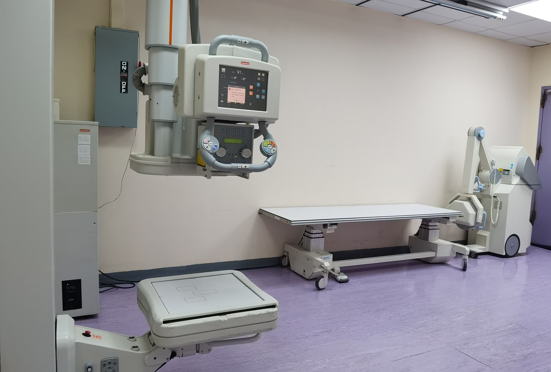
In practice, radiology technicians adjust the machine’s settings based on the body part being examined and the patient’s size. For pregnant patients, many facilities have a “pregnancy protocol” that automatically lowers the kilovoltage and milliamperage, further cutting the dose while preserving diagnostic quality. The combination of these technical safeguards and physical shielding is what keeps the radiation exposure well within safe limits. X-rays can come in various views—such as postero-anterior (PA), lateral, or oblique—each offering a slightly different perspective to aid diagnosis. The radiologist, a medical doctor specializing in imaging interpretation, plays a crucial role in ensuring the images are clear and accurately read, minimizing the need for repeat scans.
Is it safe to get an X‑ray while pregnant?
C
urrent guidance from the American College of Obstetricians and Gynecologists (ACOG) states that a single, properly shielded X‑ray is permissible when the diagnostic benefit is clear. The UK’s National Health Service (NHS) echoes this, noting that “the radiation dose from most diagnostic X‑rays is far below the level that could cause fetal harm.” The U.S. Food and Drug Administration (FDA) classifies diagnostic X‑ray devices as “generally safe” for pregnant patients, provided that the lowest‑possible dose is used and shielding is applied. This consensus from leading health authorities should offer significant reassurance.
Risk arises mainly from repeated exposures or high‑dose procedures (such as a full‑body CT scan). The CDC’s Radiation Emergency Management guidelines suggest keeping the cumulative fetal dose under 5 mGy (approximately 5 mSv) during the entire pregnancy—a threshold well above the dose from a typical chest X‑ray (0.02 mSv). Therefore, occasional imaging for urgent medical reasons is usually fine, but elective X‑rays should be postponed or replaced with non‑radiating alternatives when possible. It's important to understand that the historical concerns about radiation in pregnancy largely stemmed from much higher doses (e.g., from therapeutic radiation or older equipment) than those used in modern diagnostic imaging.
Misconceptions often center on the idea that any radiation is automatically dangerous. In reality, the dose from a standard dental or abdominal X‑ray is comparable to the radiation you encounter on a short airplane flight. The key is to ensure that the imaging is medically indicated and that protective measures are in place.
X‑ray vs. CT scan: which is safer in pregnancy?
Computed tomography (CT) scans use a series of X‑ray beams to create cross‑sectional images and typically deliver a higher dose—about 1 to 10 mSv for a chest CT, compared with 0.02 mSv for a standard chest X‑ray. Because of this higher exposure, ACOG recommends CT only when the diagnostic information cannot be obtained with a lower‑dose modality, such as MRI or ultrasound. If a CT is absolutely necessary, ask the radiology team to use a pregnancy‑specific protocol and abdominal shielding.
How does radiation from an X‑ray compare to everyday sources?
To put the numbers in perspective, a single dental bitewing X‑ray delivers roughly 0.005 mGy, which is equivalent to the natural background radiation you receive in about 10 minutes of normal daily life. A 30‑minute flight at cruising altitude adds about 0.03 mGy. Even the most common X‑ray exams remain far below the 5 mGy safety threshold set by the International Commission on Radiological Protection (ICRP). Understanding this comparison can help reduce anxiety about the “invisible” risk.
The ALARA Principle: As Low As Reasonably Achievable
When it comes to radiation exposure, the guiding principle for medical professionals is ALARA: "As Low As Reasonably Achievable." This means that every effort is made to minimize radiation dose to the patient while still obtaining the necessary diagnostic information. For pregnant patients, this principle is applied even more rigorously. It involves using the lowest possible X-ray machine settings, limiting the number of images taken, ensuring precise targeting of the area of interest to avoid unnecessary exposure to other body parts, and crucially, applying lead shielding to protect the abdomen and pelvis. You can feel confident that your radiology team is trained to follow this principle for your safety and your baby's.
Questions to ask your radiology team
Before the exam, consider asking: (1) Will a lead apron be used to shield the abdomen? (2) Is there a low‑dose or “pregnancy” protocol for this study? (3) What is the estimated fetal dose for this specific X‑ray? (4) Are there non‑radiating alternatives that could provide the same diagnostic information? Having these answers empowers you to make an informed decision and ensures the lowest possible exposure.
Safety by trimester
First trimester (organogenesis)
The first 13 weeks of pregnancy are when the baby’s major organs form, making this period the most vulnerable to teratogenic effects of high‑dose radiation. ACOG advises that X‑ray examinations in the first trimester should be limited to situations where the benefit clearly outweighs the risk, such as suspected ectopic pregnancy or severe trauma. When an X‑ray is unavoidable, lead shielding over the abdomen and a low‑dose protocol are mandatory. This "critical window" of organogenesis means that while the risk from diagnostic X-rays is still very low, medical providers are extra cautious and will only proceed if truly essential for your health or your baby's.
Because the fetus is most sensitive to radiation‑induced malformations during organogenesis, clinicians often prefer ultrasound as the first‑line imaging tool in early pregnancy. If an X‑ray cannot be avoided, the radiology department will typically calculate the exact fetal dose and discuss it with you before proceeding. In many cases, the dose remains well under the 5 mGy threshold, providing reassurance that a single necessary exam is unlikely to cause harm.
Second trimester (fetal growth)
During weeks 14‑27, the fetus’s tissues are less sensitive to radiation‑induced malformations, and the risk of growth retardation is low. Consequently, most diagnostic X‑rays (e.g., chest, extremity, dental) are considered safe with standard shielding. The NHS recommends confirming pregnancy status before any radiology order and using a lead apron whenever possible. While the risk of major malformations has significantly decreased by this stage, the developing central nervous system is still maturing, so the ALARA principle continues to be important.
Second‑trimester imaging often focuses on maternal health issues—such as evaluating a suspected pneumonia or checking a broken bone—rather than fetal assessment. Because the benefit of accurate diagnosis generally outweighs the minimal radiation risk, clinicians feel comfortable proceeding with the exam. Still, the principle of “as low as reasonably achievable” (ALARA) remains central, so the lowest‑dose settings are always selected.
Third trimester (pre‑birth preparation)
In the final trimester, the main concern shifts to potential effects on fetal growth and the risk of preterm labor from high‑dose exposures. Routine X‑rays remain low risk, but cumulative exposure should still be tracked. The FDA notes that radiation doses below 0.05 Gy (50 mGy) have not been associated with adverse fetal outcomes, a level far exceeding typical diagnostic doses. At this stage, imaging might be needed for conditions that could impact the delivery plan, such as assessing lung health or evaluating bone injuries, where the diagnostic information is crucial for safe maternal and fetal outcomes.
When a third‑trimester X‑ray is ordered, the radiology team will again verify that shielding is in place and that the machine is set to a low‑dose protocol. If the imaging is for a maternal condition that could affect delivery (such as a pulmonary embolism), the benefit of prompt diagnosis is especially important. Nevertheless, providers continue to assess whether an ultrasound or MRI could provide the same information without radiation.
What if I had an X‑ray before I knew I was pregnant?
This is a very common worry, and it's important to take a deep breath. If you had an X‑ray before you knew you were pregnant, the risk to your baby is almost certainly negligible. Most diagnostic X‑rays deliver doses far below the threshold for concern, and the "all or nothing" principle often applies in very early pregnancy: either the exposure was too low to cause harm, or it might result in early miscarriage (which often goes unnoticed) rather than birth defects. The ACOG committee opinion on radiation exposure during pregnancy specifically addresses this, stating that the risk of major malformations from typical diagnostic X-rays is extremely low.
Breastfeeding
Radiation does not pass into breast milk, and the American Academy of Pediatrics (AAP) confirms that mothers can safely continue breastfeeding after an X‑ray. No special timing or cessation is required.
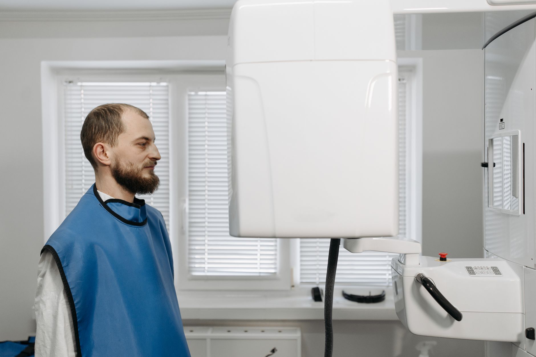
Dental X‑rays are common and can be performed safely with appropriate shielding.
How many X‑rays are safe during pregnancy?
There is no exact “number” of X‑rays that is universally safe because the important factor is cumulative radiation dose, not the count of images. The International Commission on Radiological Protection (ICRP) recommends keeping fetal exposure under 5 mGy for the entire pregnancy. A typical chest X‑ray delivers about 0.02 mGy, while a dental bitewing is roughly 0.005 mGy. This means a pregnant person could theoretically undergo dozens of low‑dose X‑rays without approaching the safety threshold, assuming proper shielding each time.
However, higher‑dose studies—such as abdominal or pelvic X‑rays—can deliver 0.1‑0.3 mGy per exam. In such cases, clinicians limit the number of examinations and may opt for alternative imaging. If you have already had multiple X‑rays early in pregnancy, discuss the cumulative dose with your provider; they can request a dosimetry report if needed. It's also worth remembering that we are all exposed to natural background radiation every day from cosmic rays, the earth, and even certain foods. The dose from a single diagnostic X-ray is often a fraction of this natural exposure.
It’s also helpful to know that the fetal dose from a single lumbar spine X‑ray (often used for back pain) is still well below the 5 mGy limit, but because the abdomen is directly in the beam’s path, extra care with shielding is essential. When multiple studies are required—for example, after a car accident—radiology departments will calculate the total dose and may prioritize the most critical exams.
Safe dosage / amount / brands
Because X‑rays are medical procedures rather than over‑the‑counter products, “brands” are not applicable. The safety focus is on the dose and shielding. Below is a quick reference for common diagnostic X‑ray types and their typical fetal dose equivalents:
Exam type
Typical fetal dose (mGy)
Shielding recommendation
Chest X‑ray (postero‑anterior)
0.02
Lead apron covering abdomen
Dental bitewing
0.005
Thyroid shield; abdominal lead apron optional
Abdominal X‑ray (single view)
0.1‑0.3
Full abdominal lead apron; consider alternative imaging
Extremity (hand/foot) X‑ray
0.001‑0.005
Usually no shielding needed for distal limbs
Mammogram
<0.01
Lead apron covering abdomen
When an X‑ray is ordered, ask the technologist whether a lead apron will be used and whether the machine is set to a “pregnancy” protocol, which reduces exposure. If you are undergoing a series of studies (e.g., multiple orthopedic X‑rays), request a cumulative dose estimate from the radiology department. Knowing the exact number helps you and your provider weigh the benefits against any theoretical risk.
In addition to shielding, some facilities employ “collimation,” a technique that narrows the X‑ray beam to the area of interest, further limiting stray radiation. Modern digital detectors also require less radiation to produce a clear image compared with older film‑based systems, meaning newer equipment is inherently safer for pregnant patients. Medical physicists play a critical role behind the scenes, ensuring that X-ray equipment is properly calibrated and doses are optimized for patient safety, especially for vulnerable populations like pregnant individuals.
Side effects and risks
For most diagnostic X‑rays, the primary risk is theoretical and relates to the tiny amount of ionizing radiation that could potentially cause DNA damage. At the low doses used in routine imaging, the absolute risk of a birth defect is estimated to be less than 1 in 10,000. This is comparable to the background risk of congenital anomalies in the general population. The type of potential damage from radiation can include cellular changes, genetic mutations, or, at very high doses, cell death. However, diagnostic X-rays are specifically designed to operate at levels far below these thresholds.
Potential short‑term side effects for the mother are rare but can include skin reddening if the beam is focused on a small area for an extended time. More concerning are cumulative high‑dose exposures, which could increase the chance of fetal growth restriction or, in extreme cases, miscarriage. If you notice unexplained bleeding, severe abdominal pain, or a sudden change in fetal movement after an X‑ray, contact your obstetric provider promptly.
It’s also worth noting that the psychological impact of worrying about radiation can be significant. Studies have shown that excessive anxiety itself can affect maternal stress levels, which may indirectly influence pregnancy outcomes. Therefore, clear communication with your care team and understanding the actual numbers can help alleviate unnecessary stress.
Safer alternatives / other safe options
Ultrasound – Uses sound waves, no radiation, and provides excellent detail for soft‑tissue evaluation, especially for abdominal or pelvic conditions and fetal monitoring.
MRI scan (non‑contrast) – Offers high‑resolution images of the brain, spine, and joints without ionizing radiation. It's often preferred for complex soft-tissue issues.
CT scan with shielding – If cross‑sectional imaging is essential, low‑dose protocols with lead shielding can limit fetal exposure, but this is a secondary option to MRI or ultrasound.
Digital tomosynthesis – A newer technique that reduces dose compared with conventional CT for certain musculoskeletal concerns.
Magnetic resonance angiography (MRA) without gadolinium – Useful for vascular assessment when radiation must be avoided. Gadolinium contrast is generally avoided in pregnancy unless absolutely critical.
Fluoroscopy with pulsed mode – When real‑time imaging is required (e.g., during certain procedures), using pulsed fluoroscopy and shielding can keep dose low.
Low‑dose chest X‑ray – If a chest X‑ray is unavoidable, request the “ALARA” setting and lead apron; the dose remains minuscule.
Physical examination and clinical assessment – Sometimes, a thorough physical exam and medical history can provide enough information to delay or avoid imaging.
When discussing alternatives, it’s helpful to ask your provider which modality will give the most diagnostic information with the least risk. For many musculoskeletal injuries, an MRI or ultrasound can replace an X‑ray entirely. In obstetric care, ultrasound is the gold standard for fetal monitoring and is completely free of ionizing radiation.
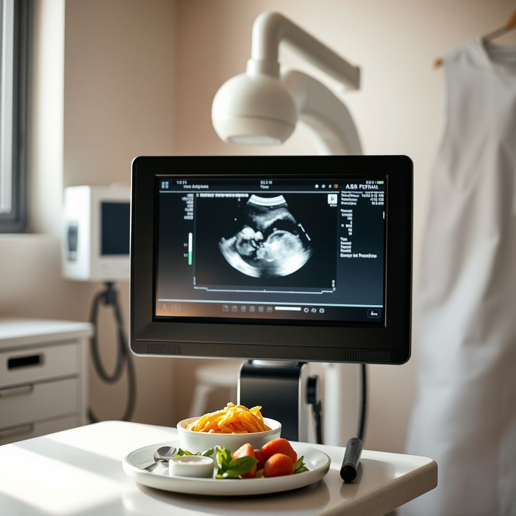
Ultrasound provides a radiation‑free alternative for many diagnostic needs during pregnancy.
Related items – safety at a glance
Imaging modality
Verdict
One‑line note
Mammogram
✅ Generally safe
Low‑dose breast imaging; lead shielding protects the fetus.
CT scan
⚠️ Use cautiously
Higher dose; only if essential and with shielding.
PET scan
❌ Best avoided
Radioactive tracer poses higher fetal exposure.
Fluoroscopy
⚠️ Use cautiously
Continuous radiation; limit duration and shield abdomen.
Radiation therapy
❌ Best avoided
Therapeutic doses are far above diagnostic levels.
DEXA scan (bone density)
⚠️ Talk to your doctor first
Low dose, but often elective; typically postponed until after pregnancy.
Myth vs. fact
Myth: Any X‑ray will cause a miscarriage.
Fact: Diagnostic X‑rays deliver doses far below the threshold known to cause miscarriage; only high‑dose therapeutic radiation poses that risk. The body's natural repair mechanisms are highly efficient at managing low-level radiation exposure.
Myth: You must stop breastfeeding after an X‑ray.
Fact: Radiation does not enter breast milk, so breastfeeding can continue as normal. There's no need to pump and dump or wait.
Myth: All X‑ray procedures are equally risky.
Fact: The risk depends on the type of exam, dose, and whether shielding is used; dental and extremity X‑rays are among the lowest‑risk, while abdominal X-rays carry a slightly higher, but still low, dose.
Myth: I can’t have an X‑ray if I’m trying to conceive.
Fact: There's no evidence that diagnostic X‑rays before conception affect fertility or future pregnancy outcomes. If you are trying to conceive and need an X-ray, it's generally safe to proceed.
Key takeaways
Most single diagnostic X‑rays are safe with lead shielding; avoid unnecessary repeats.
The first trimester is the most sensitive period; limit X‑rays to urgent situations.
Keep cumulative fetal exposure under 5 mGy; a typical chest X‑ray is only 0.02 mGy.
The ALARA principle (As Low As Reasonably Achievable) guides all X-ray procedures during pregnancy.
If you had an X-ray before knowing you were pregnant, the risk is extremely low.
Ask your provider about alternatives such as ultrasound or MRI whenever possible.
If you notice bleeding, severe pain, or changes in fetal movement after imaging, call your obstetrician right away.
Frequently asked questions
can an x-ray harm my unborn baby
In most cases, a single diagnostic X‑ray with proper shielding does not harm the unborn baby; the radiation dose is far below the level associated with birth defects. The risk is considered negligible by leading medical organizations.
what are the risks of x-ray during pregnancy
The primary risk is a very low‑level exposure to ionizing radiation, which could theoretically increase the chance of a birth defect if the dose exceeds 5 mGy, a threshold rarely reached with standard imaging. The overall risk remains extremely low.
how to minimize x-ray exposure during pregnancy
Ask for lead apron shielding, request the lowest‑dose protocol, limit the number of exams, and consider non‑radiating alternatives like ultrasound or MRI when appropriate. Always inform your healthcare team of your pregnancy status.
can i get an x-ray if i am pregnant and have a toothache
Yes—dental X‑rays are considered low‑risk and can be performed safely with a lead apron; inform the dentist of your pregnancy so they can use proper shielding. The small beam and distance from the abdomen make it very safe.
are x-rays during pregnancy covered by insurance
Most health insurance plans cover medically necessary X‑rays during pregnancy, but coverage details vary; check with your insurer for any prior‑authorization requirements. It's always a good idea to confirm coverage beforehand.
what are the guidelines for x-ray use during pregnancy
Guidelines from ACOG, NHS, and FDA advise using X‑rays only when the clinical benefit outweighs the theoretical risk, employing lead shielding, and keeping cumulative fetal dose under 5 mGy. The ALARA principle is paramount.
can an x-ray cause birth defects
Only high‑dose radiation—far above that from routine diagnostic X‑rays—has been linked to birth defects; standard X‑ray exams with shielding have not been shown to increase this risk. The estimated risk is less than 1 in 10,000.
what should i tell the radiology tech about my pregnancy
Let the technologist know you’re pregnant so they can apply a lead apron, select a low‑dose protocol, and confirm the estimated fetal dose before the exam begins. This ensures all necessary precautions are taken.
is a dental x-ray safe in the first trimester
Dental X‑rays deliver a minuscule dose (about 0.005 mGy) and are considered safe even in the first trimester when proper shielding is used; they are often the preferred imaging method for dental issues during early pregnancy.
Can X‑rays cause long‑term fertility issues for my baby?
There is no evidence to suggest that diagnostic X‑rays during pregnancy, especially at the low doses typically used, cause long‑term fertility issues for the baby later in life. Concerns about fertility are typically associated with much higher, therapeutic levels of radiation.
What if my doctor says an X‑ray is absolutely necessary?
If your doctor insists an X‑ray is absolutely necessary, it means the diagnostic information is critical for your health or your baby's, and the benefit outweighs the minimal theoretical risk. Trust your provider's judgment, but always ask about shielding and low-dose protocols.
When to call your doctor
Contact your obstetric provider immediately if you experience any of the following after an X‑ray: unexplained vaginal bleeding, severe abdominal pain, sudden loss of fetal movement, or signs of infection at the imaging site. Also call if you have any concerns about the number of X‑rays you’ve had or if you’re unsure whether shielding was used. This information is for educational purposes only and does not replace personalized medical advice.
References
American College of Obstetricians and Gynecologists. “Radiation Exposure During Pregnancy.” ACOG Committee Opinion, 2022.
National Health Service (UK). “Radiation and Pregnancy.” NHS Patient Information, 2023.
U.S. Food and Drug Administration. “Radiology Devices – Pregnancy and Radiation Safety.” FDA Guidance, 2021.
Centers for Disease Control and Prevention. “Radiation Emergency Management – Pregnancy.” CDC, 2020.
International Commission on Radiological Protection (ICRP). “Recommendations on Radiation Protection for Pregnant Women.” ICRP Publication 103, 2007.
American Academy of Pediatrics. “Breastfeeding After Radiologic Examinations.” AAP Policy Statement, 2019.
American College of Radiology (ACR). “ACR Practice Parameter for Imaging Pregnant Patients.” ACR, 2023.
Editor's pick for this topic
Not sure about the label on Is X Ray Safe During Pregnancy Trimester Risks Explained products?
Snap the ingredients list (or paste it, or scan the barcode) and SafeFilter checks every ingredient against your stage of pregnancy — flagging what to avoid, what needs care, and what's fine.
When Shubhra Mishra was expecting her first child in 2016, she was overwhelmed by conflicting food advice — one site said yes, another said never. By the time her second baby arrived in 2019, she realized millions of mothers face the same confusion.
That sparked a five-year journey through clinical nutrition papers, cultural diets, and expert conversations — all leading to BumpBites: a calm, compassionate space where science meets everyday motherhood.
Her long-term vision is to build a global community ensuring safe, supported, and free deliveriesfor every mother — because no woman should face pregnancy alone or uninformed. 🌿
🌍 Stand with mothers, shape safer guidance
Join a small circle of experts who review BumpBites articles so expecting parents everywhere can decide with confidence.