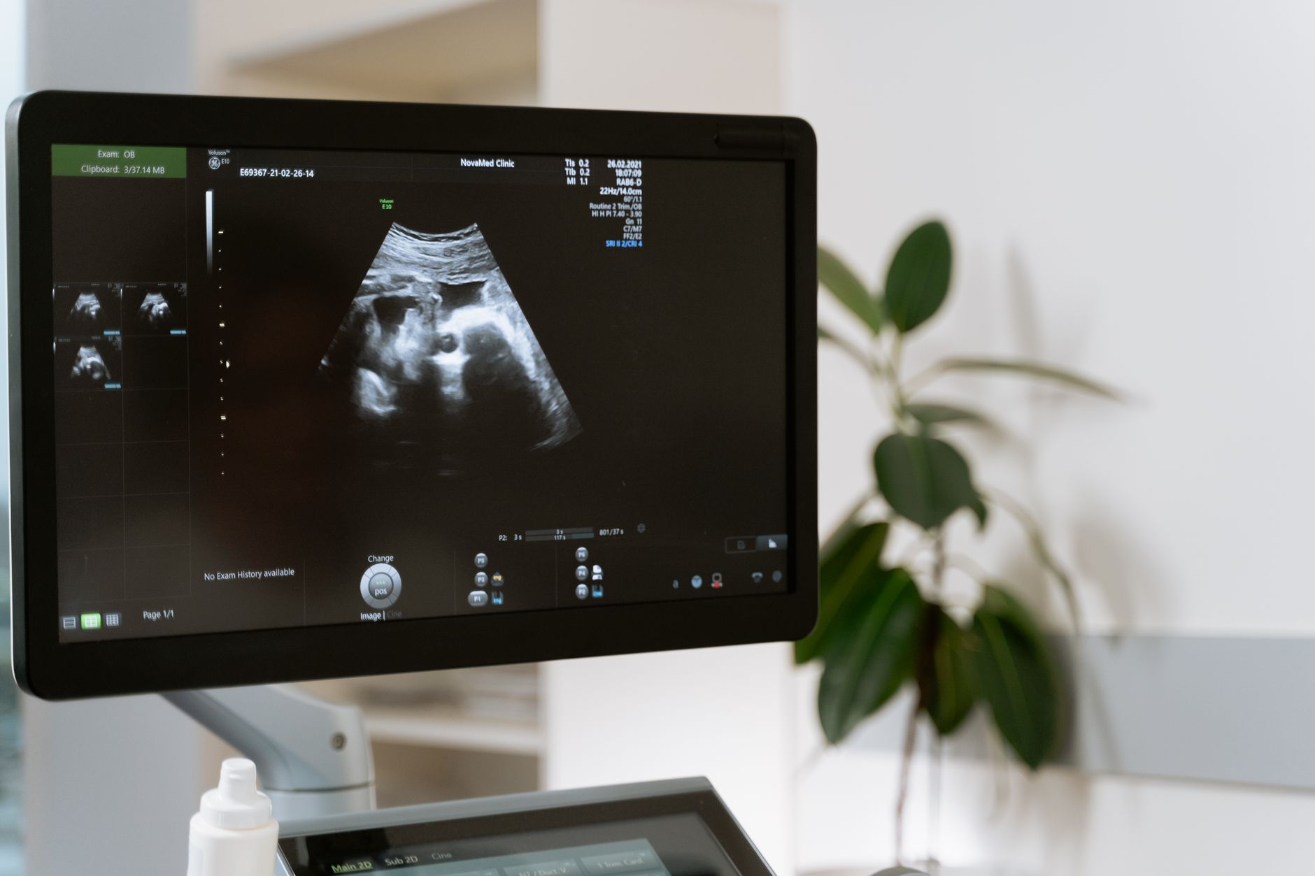
At sixteen weeks pregnant, the ultrasound reveals the baby’s head, spine, limbs, and early facial features, confirming growth, heartbeat and placenta position.
By Shubhra Mishra — a mom of two who turned her own confusion during pregnancy into BumpBites, a global mission to make food choices clear, safe, and stress-free for every expecting mother. 💛
Check whether any food is safe during pregnancy with the BumpBites Food Safety Checker.
Download the Complete Pregnancy Food Guide (10,000 Foods) 📘
Instant PDF download • No spam • Trusted by thousands of moms
💡 Your email is 100% safe — no spam ever.
Quick take: At sixteen weeks pregnant, the ultrasound lets you see your baby's head, limbs, heart, and often its gender. Measurements are usually within a normal range, the scan lasts about 20‑30 minutes, and it’s considered safe for both you and the baby.
It’s 2 a.m., you’ve just felt a flutter and are scrolling through “what does a 16 week ultrasound show.” Your heart races a little, and you wonder whether the images you’ll see are normal or if something could be wrong. You’re not alone—most first‑time parents feel the same mix of excitement and anxiety at this milestone.
In this guide we’ll walk you through exactly what happens during a sixteen‑week ultrasound, what the images mean, how big your baby should be, whether gender can be seen, and how to prepare for a smooth appointment. By the end you’ll have a clear picture of what to expect, how to interpret the report, and when to reach out to your provider.
What does a 16‑week pregnancy ultrasound show?
A sixteen‑week scan is technically an early anatomy scan. The sonographer will evaluate several key structures:
Head and brain: The skull bone is beginning to harden, and the brain’s ventricles can be measured.
Spine: A continuous line of vertebrae should be visible.
Heart: Four chambers and a beating rhythm are usually seen; a Doppler can assess blood flow.
Limbs: Arms, forearms, hands, and fingers, as well as legs, lower legs, and feet, are usually distinct.
Facial features: Eyes, nose, and mouth start to form, giving the baby a recognizable shape.
Placenta and amniotic fluid: Their location and amount are checked for any concerns.
These images give clinicians a baseline for growth and help spot major structural anomalies early enough for further testing if needed.
Because the scan is still early, some finer details—like the tiny eyelids or the exact shape of the palate—may not be crystal clear yet. That’s why the next anatomy scan at around 20 weeks often provides a more detailed view.
Extra queries answered here:
Can a 16‑week ultrasound detect heart defects? Major cardiac abnormalities, such as a missing chamber or severe valve problems, can sometimes be spotted, but many subtle defects are better evaluated later.
What does a 16‑week ultrasound report mean? The report lists measurements (head circumference, femur length, etc.), notes the presence of key structures, and flags any findings that may need follow‑up.
Typical view of a 16‑week baby during an anatomy scan.
How big is the baby at 16 weeks on ultrasound?
At sixteen weeks, the fetus is roughly the size of an avocado or a small sweet potato. On ultrasound, the crown‑rump length (CRL) usually measures about 11.5 cm (4.5 in), and the head circumference (HC) averages 11 cm (4.3 in). The femur length (FL) is typically around 1.3 cm (0.5 in).
Below is a quick reference for the normal measurement ranges you might see in a 16‑week report:
Measurement
Typical range at 16 weeks
Crown‑rump length (CRL)
10.5 – 12.5 cm
Head circumference (HC)
10.5 – 11.5 cm
Femur length (FL)
1.2 – 1.4 cm
Abdominal circumference (AC)
8.5 – 9.5 cm
These ranges reflect healthy variation; a measurement slightly outside the range does not automatically mean a problem. Growth is tracked over time, so the most important factor is whether the baby’s growth trajectory stays consistent.
Comparing the 16‑week scan to the later 20‑week scan helps illustrate how quickly the baby grows:
Week
Typical CRL
Typical HC
Typical FL
16
11.5 cm
11 cm
1.3 cm
20
16.5 cm
16 cm
2.4 cm
Seeing these numbers side by side can reassure you that the baby’s growth is on track.
Can you see gender at 16 weeks ultrasound?
Yes—most sonographers can identify the baby’s sex at sixteen weeks, but it’s not guaranteed. The genital tubercle, which later becomes either a penis or clitoris, is usually visible if the baby’s position is favorable and the equipment is high‑resolution.
Factors that affect visibility include:
Baby’s orientation (e.g., facing the probe)
Maternal body habitus (higher BMI can reduce image clarity)
Equipment quality (newer machines have better resolution)
Sonographer experience
If the gender isn’t clear, the sonographer will note “unable to determine” and may suggest a follow‑up scan later in the second trimester.
For those who prefer to keep gender a surprise, you can ask the provider to withhold that information. The report will still list the anatomical findings without mentioning sex.
Is it normal to have a small crown‑rump length at 16 weeks?
A small CRL can be a source of worry, but it’s often just a variation of normal growth. At sixteen weeks, the CRL range (10.5 – 12.5 cm) allows for a few centimeters of difference.
Reasons for a smaller measurement include:
Earlier ovulation date than estimated
Genetic factors that influence size
Maternal nutrition or hydration status at the time of the scan
Clinicians typically compare the current measurement to earlier scans. If the baby’s growth rate is steady, a single small CRL usually isn’t a red flag. However, persistent lagging growth may prompt additional monitoring, such as a follow‑up ultrasound in 2‑4 weeks or a detailed anatomy scan at 20 weeks.
In short, a slight deviation is normal; consistent lagging or accompanying concerns (like reduced amniotic fluid) warrant a conversation with your provider.
What are the common concerns during a 16‑week ultrasound?
First‑time parents often wonder about the following:
Is the baby developing normally? The sonographer checks the brain, spine, heart, and limbs for major structural issues.
What if I can’t see the gender? It’s common to need a second look; the provider can schedule a later scan if desired.
Am I safe from radiation? Ultrasound uses sound waves, not ionizing radiation, making it safe for both mother and baby.
Will the scan be painful? The transducer glides over the abdomen with a gel; you may feel mild pressure but no pain.
What if something looks off? Most findings are minor variants; if a concern arises, your provider will discuss next steps, which could include additional imaging or specialist referral.
Addressing these concerns early can reduce anxiety and help you focus on the experience.
How long does a 16‑week pregnancy ultrasound appointment last?
Typical appointments run between 20 and 30 minutes. The first 5‑10 minutes involve positioning, applying gel, and capturing basic images. The next 10‑15 minutes are dedicated to detailed anatomy views, and the final few minutes are for reviewing the images with you and answering questions.
If you’re having a more comprehensive early anatomy scan or if the sonographer needs extra time to capture a clear view of the heart or limbs, the session may stretch to 40 minutes.
To make the most of the time, bring a list of questions, your previous prenatal records, and any relevant lab results (like a positive nuchal translucency screen).
What should I expect during a 16‑week anatomy scan?
The scan begins with you lying on a comfortable exam table. A warm gel is spread over your abdomen; the gel helps the transducer move smoothly and eliminates air pockets that could distort the image.
As the sonographer slides the probe, you’ll see a monitor displaying a moving gray‑scale image. You’ll likely hear a faint “whoosh” as the machine sends and receives sound waves. The baby’s heartbeat—usually around 120‑160 beats per minute—will be audible, providing an instant reassurance.
Key moments during the scan include:
Head and brain view: The sonographer will measure the head circumference and look for the cavum septi pellucidi (a small fluid‑filled space that appears as a bright spot).
Heart assessment: Using color Doppler, the technician assesses the flow through the chambers and major vessels.
Limbs and hands: Fingers and toes become distinct; the sonographer may count them.
Spine alignment: A continuous line of vertebrae should be visible.
Gender check (optional): If you’ve requested it, the sonographer will try to capture the genital area.
After the images are captured, the sonographer will usually show you a short video loop of the baby’s movements—a 16‑week ultrasound video that many parents love to share with family.
Relax in a comfortable position—your provider will guide you through the scan.
Tips for first‑time moms:
Wear comfortable, loose‑fitting clothing that allows easy access to the abdomen.
Stay hydrated but avoid a full bladder if your provider asks you to empty it first.
Bring headphones if you prefer to hear the heartbeat privately.
Write down any questions you have before the appointment; the sonographer’s time is limited.
Are there any risks to a 16‑week ultrasound?
Ultrasound is considered a low‑risk, non‑invasive imaging modality. The World Health Organization (WHO) and the American College of Obstetricians and Gynecologists (ACOG) state that routine obstetric ultrasounds are safe when used for appropriate medical indications.
Potential, but rare, concerns include:
Thermal effects: Prolonged exposure to high‑intensity sound can raise tissue temperature, but standard scans keep the exposure well below harmful levels.
Mechanical index: Excessive acoustic power could theoretically cause micro‑cavitation, yet modern machines limit this automatically.
Because the scan is brief and follows established safety guidelines, there is no evidence linking a single 16‑week ultrasound to adverse outcomes.
If you have specific concerns—such as a previous miscarriage, a high‑risk pregnancy, or a personal preference to limit imaging—discuss them with your provider. They can adjust the scan duration or use a “limited” protocol if needed.
From our medical team: A sixteen‑week ultrasound is a routine part of prenatal care. It provides valuable information about your baby’s growth and anatomy while posing no known risk when performed by a qualified professional. If any findings raise questions, we’ll guide you through the next steps with clear, evidence‑based recommendations.
Myth vs. fact
Myth: You can’t see the baby’s gender at sixteen weeks.
Fact: The genital tubercle is often visible at sixteen weeks, allowing gender identification in most cases, though positioning can affect visibility.
Myth: Ultrasound radiation harms the baby.
Fact: Ultrasound uses sound waves, not ionizing radiation, and is considered safe when used appropriately.
Myth: A small crown‑rump length always means a problem.
Fact: Small measurements can be within normal variation; persistent growth lag is what clinicians monitor.
Key takeaways
At sixteen weeks, the ultrasound checks head, spine, heart, limbs, and placenta for major structural development.
Typical measurements: CRL ≈ 11.5 cm, HC ≈ 11 cm, FL ≈ 1.3 cm; ranges vary, and slight differences are normal.
Gender can often be seen, but a clear view depends on baby’s position and equipment.
The appointment lasts about 20‑30 minutes; bring a question list and comfortable clothing.
Ultrasound is safe—no ionizing radiation and minimal thermal risk when performed by trained staff.
If the report notes any concerns, follow‑up imaging or specialist referral is the usual next step.
Frequently asked questions
Can you see the baby's gender at 16 weeks?
Yes—most sonographers can identify gender at sixteen weeks if the baby’s genital tubercle is clearly visible; otherwise they may note “unable to determine.”
How big is a 16‑week fetus on ultrasound?
The fetus is typically about 11.5 cm long (crown‑rump length) and weighs roughly 100 g, comparable to an avocado.
What should I expect during a 16‑week anatomy scan?
Expect a brief, gentle exam with gel applied to your abdomen, a moving image of your baby’s head, heart, limbs, and possibly gender, lasting 20‑30 minutes.
Is a 16‑week ultrasound safe for the baby?
Yes—ultrasound uses non‑ionizing sound waves and is endorsed as safe by ACOG and WHO when performed for appropriate medical reasons.
How long does a 16‑week ultrasound take?
The scan usually takes 20‑30 minutes, though complex cases may extend to 40 minutes.
What abnormalities can be detected at 16 weeks?
Major concerns such as severe heart defects, spinal abnormalities, markedly abnormal head size, or missing limbs can be seen; subtle issues often require later scans.
When to call your doctor
If you notice any of the following, contact your provider right away: severe abdominal pain, heavy bleeding, sudden loss of fetal movement, fever over 38°C (100.4°F), or if the ultrasound report mentions a serious anomaly.
This article provides general information and is not a substitute for personalized medical advice. Always discuss any concerns with your obstetrician, midwife, or qualified healthcare professional.
References
American College of Obstetricians and Gynecologists (ACOG). “Ultrasound in Pregnancy.” 2023 clinical guidance.
World Health Organization (WHO). “Recommendations on Antenatal Care for a Positive Pregnancy Experience.” 2022.
National Institute for Health and Care Excellence (NICE). “Ultrasound scanning in pregnancy.” Updated 2023.
Society for Maternal-Fetal Medicine (SMFM). “Guidelines for the Use of Ultrasound in Pregnancy.” 2021.
Royal College of Obstetricians and Gynaecologists (RCOG). “The 20‑Week Scan.” 2022.
Mayo Clinic. “Ultrasound during pregnancy: What to expect.” Accessed July 2026.
Editor's pick for this topic
Not sure about the label on 16 Week Pregnancy Ultrasound What You Ll See On The Scan products?
Snap the ingredients list (or paste it, or scan the barcode) and SafeFilter checks every ingredient against your stage of pregnancy — flagging what to avoid, what needs care, and what's fine.
When Shubhra Mishra was expecting her first child in 2016, she was overwhelmed by conflicting food advice — one site said yes, another said never. By the time her second baby arrived in 2019, she realized millions of mothers face the same confusion.
That sparked a five-year journey through clinical nutrition papers, cultural diets, and expert conversations — all leading to BumpBites: a calm, compassionate space where science meets everyday motherhood.
Her long-term vision is to build a global community ensuring safe, supported, and free deliveriesfor every mother — because no woman should face pregnancy alone or uninformed. 🌿
🌍 Stand with mothers, shape safer guidance
Join a small circle of experts who review BumpBites articles so expecting parents everywhere can decide with confidence.